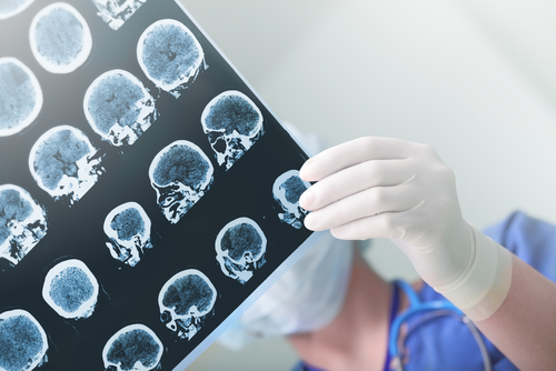
Researchers investigated the role of brain dystrophin, a protein with an important role in the stability of central nervous system (CNS) cells and neurotransmitters, in temporal lobe epilepsy and found that the protein is over-expressed in certain areas of the CNS, hinting at a compensatory mechanism in the chronic epileptic human brain.
The research paper, “Dystrophin Distribution and Expression in Human and Experimental Temporal Lobe Epilepsy,” was published in frontiers in Cellular Neuroscience.
Mutations in the dystrophin gene lead to the absence of this protein, ultimately resulting in the neuromuscular disease known as Duchenne muscular dystrophy (DMD). Increasing evience suggests that DMD is accompanied by such comorbidities as non-progressive cognitive and behavioral difficulties, and epilepsy. These brain abnormalities have been attributed to the lack of dystrophin in the CNS, where it plays an important role in the correct functioning and distribution of neurotrnasmitter receptors, and ion- and water-channels in the cell membrane.
Researchers investigated the possible role of dystrophin in neuronal excitability. They studied brain dystrophin distribution and expression in both human and in experimental models (rats) of temporal lobe epilepsy (TLE) and non-epileptic controls, focusing on the hippocampus and cerebellum, the two major brain structures containing dystrophin. While the hippocampus plays a major role in the pathogenesis of TLE, the cerebellum may be involved in anti-epileptic neuromodulation.
Results showed no differences in dystrophin expression levels and distribution in the rat models of epilepsy and the control animals. But a specific type of dystrophin, present in the hippocampus, was found at 60 percent higher levels in human TLE patients than in controls.
This difference between the human and animal experiments is due to the fact that while human TLE is characterized by the presence of neuronal cell loss, the animal model does not present neurodegeneration, the researchers said. They believe the higher levels of dystrophin may indicate “multiple potential compensatory mechanisms that could restore the balance between excitation and inhibition in brains that are prone to hyperexcitation.”
“Future studies should address how the different brain dystrophin isoforms are related to hyperexcitation, in what direction this relationship may be established and, ultimately, whether dystrophin may represent a novel target for seizure treatment,” the researchers concluded.


